The sacroiliac (SI) joints are critical yet often overlooked structures that connect the sacrum at the base of the spine to the ilia of the pelvis. In women, these joints bear unique biomechanical demands due to a wider pelvic structure, greater inherent mobility, and hormonal influences. When the SI joint becomes misaligned or dysfunctional whether through hypermobility, hypomobility, or inflammation it can trigger a cascade of compensatory changes down the open kinematic chain, frequently manifesting as altered knee angles also knows as Q-angle and persistent knee pain.
This scientific blog post explores the anatomy and biomechanics specific to females, the mechanisms linking SI joint issues to knee problems, and the evidence supporting chiropractic intervention. Anatomy and Sex Specific Biomechanics of the SI Joint. The SI joints are synovial joints with irregular, auricular-shaped surfaces designed primarily for stability and load transfer rather than large ranges of motion. Normal motion is minimal: typically less than 4° of rotation and about 1.6 mm of translation. They function as shock absorbers, transmitting forces from the upper body to the lower extremities while resisting shear.
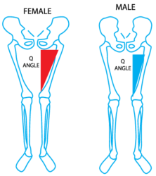
Women exhibit greater SI joint mobility than men maximum range of motion around 2.8° versus 1.2° in males. This increased laxity stems from a broader pelvis (adapted for childbirth), more pronounced ligamentous flexibility, and hormonal factors like relaxin, which softens ligaments during pregnancy. These adaptations, while essential for reproduction, predispose women to higher stresses, loads, and ligament strains at the SI joint. SI joint dysfunction (SIJD) involves aberrant motion or position either excessive movement (instability/hypermobility) or restricted movement (fixation/hypomobility). Common contributors in women include pregnancy and postpartum changes, leg length discrepancies, poor posture, repetitive stress, trauma, and muscle imbalances. Prevalence data suggest SIJD accounts for 15–30% of low back pain cases, with higher rates in females, particularly during reproductive years.
How SI Joint Misalignment Affects Pelvic Position and Knee Biomechanics.
A misaligned or dysfunctional SI joint disrupts pelvic symmetry and orientation. Common patterns include unilateral anterior or posterior pelvic tilt (torsion), iliac rotation, or sacral shear. These changes alter the position of the acetabulum (hip socket), affecting femoral alignment and tracking.
Down the kinematic chain:
- Altered hip mechanics: Pelvic torsion can cause relative hip internal or external rotation, changing the Q-angle (quadriceps angle) at the knee. Increased dynamic knee valgus, where the knee collapses inward is a frequent compensation. Studies link anterior pelvic tilt and pelvic asymmetries to greater knee valgus stress.
- Gait and loading changes: Research on female runners with SI joint pain shows reduced knee flexion, greater tibial overstride, and increased ankle dorsiflexion compared to controls. These adaptations increase ground reaction forces and uneven loading across the knee joint.
- Muscle inhibition and imbalances: SIJD often inhibits gluteal and core muscles, leading to over-reliance on quadriceps or altered patellar tracking. One study demonstrated that SI joint manipulation increased knee-extensor moment and reduced muscle inhibition.
These biomechanical shifts contribute to conditions like patellofemoral pain syndrome (PFPS), iliotibial band syndrome, and accelerated joint wear. Knee pain from SIJD may be referred (radiating to the posterior thigh or knee without true sciatica) or mechanical, often felt as aching, sharpness with stairs or transitions, or a sense of instability/buckling.
Pelvic misalignment can functionally create a leg length discrepancy, forcing compensatory pronation or supination at the foot and valgus/varus stress at the knee. In women, the combination of wider Q-angles and pelvic laxity amplifies these effects.
Clinical Presentation in Women.
Symptoms often include one-sided low back or buttock pain radiating to the groin, thigh, or knee. Pain worsens with prolonged standing, walking, single-leg activities, or rising from sitting. Pregnancy-related SIJD is especially common due to relaxin-induced laxity plus increased load, with many women experiencing persistent postpartum issues if not addressed.
Diagnosis relies on clinical tests (e.g., FABER, Gaenslen’s, Fortin’s finger test, sacral thrust) rather than imaging alone, as X-rays or MRI may miss subtle dysfunction. Differential diagnosis must rule out lumbar radiculopathy, hip pathology, or inflammatory conditions. How Chiropractic Care Addresses SIJD and Associated Knee Pain. Chiropractic care targets the root biomechanical dysfunction through precise adjustments, soft tissue techniques, and rehabilitation offering a conservative, evidence-informed approach.
High-velocity, low-amplitude (HVLA) manipulations: Side-posture or prone adjustments to the SI joint and lumbar spine restore motion in hypomobile segments, reduce pain, and improve load distribution. A study of 32 women with SIJD found greater improvements in pain and mobility (immediate, 2 days, and 30 days post-treatment) with HVLA to the SI and lumbar regions versus other therapies.
- Mobilizations and muscle energy techniques (MET): Gentler options suit hypermobile or acute cases, helping normalize joint play.
- Full kinetic chain assessment and correction: Addressing lumbar, hip, foot, or leg length issues prevents recurrence. Adjustments can quickly reduce quadriceps inhibition, improving knee function.
- Rehabilitative exercises: Core stabilization (e.g., planks, bird-dogs), glute strengthening, pelvic tilts, and proprioceptive training enhance force closure of the SI joint. Pelvic belts provide temporary stability, especially postpartum.
Randomized trials support chiropractic manipulation for SIJD, showing reductions in pain and disability comparable to or faster than exercise alone. One trial comparing manipulation, MET, and home exercise found manipulation provided the quickest relief.
Outcomes for knee pain: By restoring pelvic symmetry and hip mechanics, chiropractic reduces abnormal knee stresses. Patients often report decreased referred knee pain and improved gait stability. Multimodal care (adjustments + exercise) yields the best long-term results.
Limitations and Integrated Care.
While highly effective for many, results vary. Hypermobile patients may need more emphasis on stabilization than manipulation. Severe cases or those with neurological deficits warrant medical co-management, possibly including injections or, rarely, fusion. Chiropractic is not a standalone cure but excels in conservative management.
Prevention and Long-Term Management
Maintain strong core and glute muscles, practice good posture, use proper lifting mechanics, and consider supportive footwear. Postpartum screening for pelvic stability is crucial. Regular chiropractic check ups can catch subtle dysfunctions early.
Conclusion
In women, SI joint misalignment is a common but under-recognized driver of knee angle alterations and pain via disrupted pelvic and lower limb biomechanics. Chiropractic care, grounded in restoring alignment, mobility, and neuromuscular control, offers significant relief and functional improvement. Early intervention can break the pain cycle and prevent chronic issues.
If you experience pelvic, low back, or knee pain that hasn’t responded to isolated knee treatments, consult a qualified chiropractor for a comprehensive biomechanical evaluation.